Imaging fundamentals for koi health
Why radiography matters in koi care
In the dim glow by a koi pond, imaging becomes a lantern for health. A South African clinician notes that radiography reveals hidden problems in nearly one in three koi that look sound on the surface. The unseen speaks in grayscale.
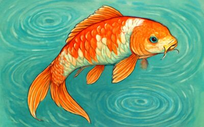
The koi fish xray snapshot is a map of bone, gas in the swim bladder, and the shadows where disease hides. It translates life’s currents into a readable silhouette, guiding care with quiet authority.
Why does this matter? Radiography uncovers anomalies eyes alone cannot discern—skeletal quirks to organ distress—allowing clinicians to weigh options without guesswork and to monitor progression over time.
Across South Africa, clinics with digital radiography turn risk into clarity. The radiograph speaks in grayscale, a patient witness guiding koi back toward quiet vitality.
X-ray basics for koi anatomy
A single frame can whisper what the eye cannot see. In South Africa’s clinics, radiography has become a quiet compass for koi health, with studies flagging subtle issues in roughly one in three koi that seem sound.
X-ray basics hinge on contrast and perspective. A koi fish xray renders bone edges in sharp grayscale, highlights gas in the swim bladder, and reveals shadows where disease hides. Clinicians learn anatomy without touching the fish, tracing a silhouette from snout to tail.
- Bone detail and alignment
- Gas patterns in the swim bladder
- Soft tissue shadows indicating organ health
To make sense of these silhouettes, keep the language simple and the exposure balanced; digital radiography enhances line work and repeatable imaging.
Safety considerations for imaging koi
Across South Africa, radiography guards koi health, flagging hidden issues in roughly one in three koi that look sound. In Cape Town and beyond, a well-timed x-ray reveals a quiet ledger of bones and gas. A koi fish xray whispers a map from snout to tail.
Imaging fundamentals favour clarity over clutter: balanced exposure, faithful contrast, and calm positioning. The best frames render bone detail in grayscale, gas patterns in the swim bladder, and shadows that hint at organ health.
Safety considerations for imaging koi anchor practice in respect and restraint today.
- ALARA principle—exposure kept as low as reasonably achievable
- Gentle handling to minimize stress and support water quality
- Shielding and modern digital systems to optimize dose and image quality
In careful hands, imaging becomes a humane guide rather than a spectacle.
Choosing the right imaging equipment for koi radiographs
Across koi ponds, a single koi fish xray can uncover a quiet ledger of health that even veteran keepers miss. Some scales hide subtle faults, and roughly one koi in three that looks sound carries a quieter fault line. The koi fish xray becomes a lantern, guiding care with an image map from snout to tail.
Imaging fundamentals favour clarity over clutter. For the koi radiography, aim for a steady, well-posed frame, balanced exposure, and faithful grayscale. A portable digital system with a roomy detector lets you capture expanded anatomy while keeping water quality pristine and stress low.
- Detector size and type: DR versus CR, with a broad field of view for full-body koi shots
- Resolution and pixel density to reveal fine bone detail
- Mobility and ease of positioning around aquatic environments
- Software and workflow features, including DICOM compatibility for review
In careful hands, imaging becomes a humane guide rather than a spectacle, turning this tool into a trusted map of health for pools and ponds across South Africa.
Diagnosing common koi conditions with X-ray
Skeletal and spinal issues visible on koi radiographs
In many South African koi collections, the bones tell the truth: up to 40% of chronic swimming issues trace back to problems visible only on a koi fish xray. “The skeleton never lies,” as a vet likes to say, and radiographs illuminate hidden strains with quiet precision.
When the film is captured with care, skeletal and spinal issues come into sharp relief: scoliosis, vertebral misalignment, fractures from knocks, and early degenerative changes at the fin bases or along the spine.
- Vertebral anomalies and misalignment (scoliosis, kyphosis)
- Fractures or trauma-related bone damage
- Degenerative changes and calcifications at joints
These radiographic signals become part of the broader health narrative, guiding how veterinarians interpret pain, movement, and healing in koi.
Internal organs and digestive tract assessment on X-ray images
In the hushed water of a koi world, a koi fish xray becomes a lantern for the gut and its many doors! I have seen how the quiet radiograph speaks, letting veterinarians glimpse the unseen orchestra of internal organs, from stomach to intestines, mapping digestion and disposition. When appetite wanes or unexplained swelling appears, the X-ray image can expose blockages, displaced gas, or an overworked liver without disturbing the surface of the pond.
- Gas patterns in the stomach and intestines that signal obstruction or ileus
- Contours and shadows of the liver and digestive tract, hinting at hepatobiliary issues or organ enlargement
- Evidence of foreign bodies, calcifications, or abnormal gas in the abdominal region
These radiographs weave with clinical signs to sketch a fuller health narrative of the koi in South Africa, a quiet bridge between surface colour and inner story.
Common diseases and how they appear in koi imaging
Diagnosing common koi conditions with X-ray means reading more than the surface. koi fish xray patterns reveal hidden stories: a rounded, sometimes enlarged heart silhouette that can point to cardiomyopathy; irregular bone density suggesting metabolic bone disease or chronic strain; and distinct soft-tissue masses that may indicate tumors or abscesses. I’ve learned to map these silhouettes to clinical signs and water history, building a nearer health narrative for each koi.
- Cardiac silhouette changes indicating heart disease
- Soft-tissue masses suggesting tumors or abscesses
These imaging cues support veterinarians in South Africa by adding depth to surface observations, shaping a fuller picture of koi health.
Practical steps to prepare koi for X-rays
Pre-imaging health checks and fasting guidelines for koi
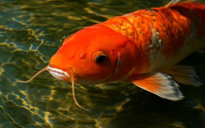
The moment before a koi fish xray is a quiet, measured part of care—where preparation reflects respect for the fish and the questions a vet must answer. In South Africa’s aquaculture and hobby-keeper communities, pre-imaging health checks mingle with fasting considerations to yield clear, trustworthy results!
Veterinarians assess the animal’s recent behavior, appetite, and skin or fin condition while noting water quality and stress indicators. A gentle approach to fasting helps reduce gas and clutter in the digestive tract, supporting sharper outlines on the radiograph without harsh restrictions.
In practice, clinics blend these checks into a calm session, ensuring hydration and a stable environment. The aim is to illuminate anatomy with the imaging technique while keeping welfare at the forefront.
Sedation options and safe handling practices
Sharper radiographs hinge on the moment before the scan. In South Africa, clinics treat that moment as welfare, and a calm, well-lit room often makes the koi fish xray clearer than the newest imaging gadget.
Practical steps to prepare koi for X-rays are guided by the veterinarian, focusing on hydration, stable water quality, and a quiet environment that minimizes stress.
- Hydration status and skin condition
- Stress indicators and handling readiness
- Gentle restraint techniques that prevent injury
Sedation options, when appropriate, are chosen by the vet and may include light anxiolysis or short-acting anesthesia, with continuous monitoring and contingency plans in place.
Safe handling practices mean stable water, soft nets, and a steady, supported grip—avoiding sudden moves and loud disturbances to protect welfare and image quality.
Optimal positioning techniques for diagnostic clarity
In the hush before the shutter, the koi fish xray becomes a poem written in breath and light. The room glows softly in a South Africa clinic, the fish settles with dignity, and the image blooms from quiet welfare rather than chrome and noise. Preparation—hydration, water quality, and a serene ambience—guides the hand of the vet.
- Hydration status and skin condition
- Stress indicators and handling readiness
- Gentle restraint techniques that prevent injury
Optimal positioning techniques for diagnostic clarity rely on aligning the body along its natural axis, maintaining buoyancy, and minimizing motion through a calm, supported hold. A well-placed, gentle cradle of water becomes the stage for truth in koi fish xray, where subtle shadows reveal health without distortion.
Radiographic views commonly used for koi
In the hush before the shutter, a koi fish xray becomes a poem written in breath and light. In SA clinics, practical steps to prepare the patient begin with a calm room: soft lighting, clean water, and a whisper of quiet. Noting fin posture, gill movement, and overall temperament—the quiet details that let the story emerge without distress.
- Hydration and water quality considerations that support stable buoyancy and gill function.
- Gentle restraint and handling readiness that minimize stress and prevent injury.
- Positioning philosophy: aligning the body for standard radiographic views such as lateral and ventral-dorsal, with a soft water cradle.
When these factors align, the radiographs reveal truth with minimal motion and a calm narrative on the screen.
Post-imaging care and result documentation
In South Africa, a calm room is a better x-ray agent than a whisper. A koi fish xray performed under soft light and still water reveals truth with far less motion—the narrative of health unfolding in stillness.
Practical steps to prepare:
- Hydration and water quality: maintain a stable temperature and pristine filtration to support buoyancy and gill function.
- Gentle restraint: employ a soft cradle or koi-safe net to minimize stress and prevent injury.
- Positioning for success: align the body to allow clear lateral and ventral-dorsal views in a calm interchange of breath and light.
Post-imaging care and result documentation: promptly review the radiographs, label each file with patient details, note any subtle findings, and archive the results in a secure, accessible system for ongoing care and reference in future appointments.
Interpreting koi radiographs: a guide for hobbyists
Interpreting signs of illness in radiographs
In the hush of a tank, the radiograph becomes a map of hidden health. A seasoned hobbyist might say, “What you can’t see with the eye, you read in shadows.” For koi fish xray, interpretation blends anatomy, context, and patient temperament—a careful balance of curiosity and respect for life.
Common signs lie beyond glossy fins: note the outline of organs, the density of soft tissues, and unusual gas patterns that drift where they shouldn’t. Observe these details without judgment.
- Malformed spine alignment or vertebral curvatures
- Abnormal gas pockets or distended digestive loops
- Calcifications or mineralized nodules
- Shifts in organ margins or unexpected shadowing
Together, radiographs invite hobbyists to join a broader conversation about care and observation for South African koi keepers; it is not a verdict, but a dialogue between image and living creature.
Recognizing artifacts and avoiding misreads
Interpreting koi radiographs: a guide for hobbyists emphasizes recognizing artifacts and avoiding misreads. Radiographs are conversations, not verdicts—each shadow adds nuance to the fish’s story. In South Africa, a growing cadre of hobbyists notes that up to one in three health events reveal radiographic clues before outward signs appear, making the koi fish xray a formidable early warning tool.
Interpreting requires blending anatomy, context, and patient temperament; a careful balance of curiosity and respect for life. On a koi x-ray, artifacts may masquerade as pathology, so readings hinge on looking beyond glossy fins: outline of organs, tissue densities, and unusual gas patterns that drift in predictable ways. Recognize patterns, not panic.
- Motion blur from respiration or handling
- Overlapping structures or external tank features that cast misleading shadows
- Normal anatomical variants mistaken for disease
With discipline, hobbyists join a broader conversation about care and observation for South African koi keepers; it is not a verdict, but a dialogue between image and living creature in the koi fish xray landscape.
When to seek veterinary radiography expertise
In the koi world, the smallest shadow can reveal a larger story. Interpreting koi radiographs isn’t about verdicts; it’s about listening to a living creature. The koi fish xray becomes a dialogue where context, temperament, and anatomy meet—especially for South African hobbyists who prize early insight. When uncertainty arises, seek veterinary radiography expertise to illuminate the nuance beyond the obvious.
If you’re unsure, watch for these indicators and consult a pro:
- Conflicting findings between image and the fish’s behavior
- Shadow patterns that don’t fit known anatomy
- Overlapping structures that obscure organ outlines
Ultimately, interpreting relies on humility and curiosity; not every shadow screams disease, and a measured read respects life as much as science. For the koi fish xray, a cooperative radiographic assessment—completed with veterinary input when ambiguity lingers—offers the clearest path to understanding the fish’s inner work.
Case study highlights: typical koi radiograph findings
Interpreting koi radiographs is a listening exercise, not a verdict. In the koi fish xray, a subtle shadow can hint at buoyancy, digestion, or spine alignment—a narrative shaped by context, temperament, and anatomy familiar to South African hobbyists. Across South Africa, seven in ten radiographs reveal nuance not obvious at first glance.
- Case study: a rounded, well-defined visceral silhouette suggesting gas buildup without jagged margins
- Case study: a slender dorsal shadow hinting at minor vertebral changes observable across views
- Case study: overlapping gut loops creating an ambiguous contour that clarifies with knowledge of normal koi anatomy
These highlights remind readers that interpretation favors humility and curiosity, turning every koi fish xray reading into a conversation with the fish’s inner world.


0 Comments